No, I just guessed that a tetraploid pollinated by a diploid would probably yield a triploid.
Some further info on “blue” roses, this time derived from varieties that “blue” with age. I haven’t read the whole series (seven so far), but I wanted to report them here for anyone else who might be interested.
The first link leads to I, II and VI. The other reports have separate links, except for III, which I cannot find.
Bot. Mag. Tokyo 83(985): 233-236 (Oct 2006)
Studies on “Bluing Effect” in the Petals of Red Rose,
I. Some Cytochemical Observations on Epidermal Cells Having a Bluish Tinge
Hitoshi Yasuda
Cytologia 39: 107-112, 1974
Studies on “Bluing Effect” in the Petals of Red Rose,
II. Observation on the development of the tannin body in the upper epidermal cells of bluing petals
Hitoshi Yasuda
Jour. Fac. Sci., Shinshu Univ. 8(1): 91-94. (1973)
Studies on “Bluing Effect” in the Petals of Red Rose,
III. The histochemical detection of iron in the bluing petals of rose.
Hitoshi Yasuda
Jour. Fac. Sci., Shinshu University 11(1): 41-46 (June 1976)
Studies on “Bluing Effect” in the Petals of Red Rose,
IV. Calcium in the blue spherical body.
Hitoshi Yasuda
Jour. Fac. Sci., Shinshu University 13(1): 79-86 (June 1978)
Studies on “Bluing Effect” in the Petals of Red Rose,
V. A Survey of the Various Bluing Types
Hitoshi Yasuda and M. Kikuchi
Cytologia 47: 717-723 (1982)
Studies on “Bluing Effect” in the Petals of Red Rose,
Vl. Further observations on the development of blue color of the spherule
Hitoshi Yasuda
Jour. Fac. Sci., Shinshu University 20(1): 15-20 (1985)
Studies on “Bluing Effect” in the Petals of Red Rose,
VII. Cytological Observation on the Epidermal Cells of Bluing Petals Incorporated into the Miscellaneous-type.
Hitoshi Yasuda and Akio Yoneda
Jour. Fac. Sci., Shinshu University 13(1): 79-86 (June 1978)
V. A Survey of the Various Bluing Types
Hitoshi Yasuda and M. Kikuchi
One hundred and forty five rose cultivars, including old roses, were used to determine the various bluing patterns of red rose petals. The upper epidermis of fresh petals, which had exhibited the bluing phenomenon, were peeled and examined microscopically.
The bluing patterns observed were grouped into three types. The first was the cell sap-type where the central vacuole of upper epidermal cells was uniformly blue without any apparent blue structures. The second was a tannin body-type in which blue spherical tannin bodies appeared in the vacuoles. The third was a miscellaneous-type which included blue structures in the vacuole other than tannin bodies.
Old roses derived from > Rosa chinensis > usually exhibited the cell sap-type of bluing while those derived from > Rosa gallica > had a tendency to exhibit a combination of the tannin body-miscellaneous bluing types. Combinations of two or even all three of the bluing types were found in some cultivars. With the long history of rose breeding, the various bluing types and their combination could have evolved through the segregation and recombination of the bluing factors, specifically when > Rosa chinensis > or > Rosa gallica > were used as parents.
The non-tannin structures may correspond to AVIs, which are found in some old roses that have no known or suspected China rose ancestry: e.g., ‘L’Evêque’ and ‘Bleu Magenta’). The tannin types might be the cultivars that are less blue in dry weather/soil, such as ‘Erinnerung an Brod’ and ‘Veilchenblau’. The blue sap type (Mr. Bluebird?) deserves further attention. Maybe a different sort of co-pigment effect that acts in solution.
More of Yasuda’s related research:
Studies on the insoluble states of anthocyanin in rose petals
Hitoshi Yasuda
Jour. Fac. Sci. Shinshu Univ. 9: 63-69 (1974)
I. The insoluble state of anthocyanin and its relationship to petal color, together with a new instance of this relationship.
Abstract
The upper epidermal cells of certain cultivars of black roses, e.g. Charles Mallerin, Josephine Bruce, Bonne Nuit and so on, occasionally hold in the cell vacuoles massive structures colored purple or purplish-red.It was observed histochemically that these massive structures showed a color change from either purplish-red or from purple to red with dilute hydrochloric acid and to bluish purple with a weak solution of sodium hydroxide. From these results the massive structure found in the red rose petals can be regarded as the insoluble state of anthocyanin.
In the surface view of those petals having these massive structures in their upper epidermal cells, the numerous black spots, which are considered to be shadows of epidermal cells, are visible even in petals whose height/width ratio of the upper epidermal cells are within the range of the red petal ones, It can therefore be said that the insoluble state of anthocyanin present in the red rose petals may play an important role in the blackening effect on the petal color.
Introduction
It is generally known that anthocyanins are present in solution in the cell sap. However, it is considered to be a rare occurrence when anthocyanin appears in insoluble states in plant cells or in tissues. According to BLANK’S review), the insoluble states of anthocyanin can be classified by the following three types:(A) a crystallized type in cell plasma (e.g > Allium> ) and in sap (e.g., juice of the blood orange).
(B) a stored type in the cell wall (e.g. > Sphagnum, Marchantia, Preissia > etc.).
(C) an “anthocyanophore” type in the cell vacuole (e.g. > Erythrea, Fuchsia, Iris, Dianthus, Delphinium, Pulmonaria > and so on).
Very recently, the present author reported that the cell vacuole of the bluing petals of the red rose, in some cases, includes anthocyanin as a component of the blue spherule, the basis of which may principally consist of a tannin-like substance). This example could be considered as another type of the insoluble state of anthocyanin.
As stated above, several observations have been made on the insoluble states of anthocyanins, but there has been relatively little information concerning their cytological and physiological investigations.
As a beginning step in detailed investigations on the insoluble state of anthocyanin, especially of the anthocyanophore type, the present paper is concerned with its significance relative to the petal color, thus providing a new example found in some petals of the so called “black rose”.
Cytologia 41: 487-492 (1976)
II. Histochemical observation on its basal substance.
In the previous paper (Yasuda 1974), it was reported that the blackish petals of certain rose cultivars (e. g. Charles Mallerin, Bonne Nuit and Josephine Bruce) occasionally include an insoluble state of anthocyanin in the central vacuoles of the upper epidermal cells, and it was also pointed out that this state contributes toward the elucidation for the presence of blackish tone to the petal color of these cultivars. In the same paper, it was described how this insoluble state of anthocyanin showed a closely similar appearance to the anthocyanophore which had been named by Weber (1934, 1936), especially to that found in > Erythraea > reported by Lippmaa (1926).
If this insoluble state of anthocyanin is analogous to the anthocyanophore, it should then be something of a massive structure composed of basal substances associated with anthocyanin. Hence, it will be necessary for the moment to attack the question of whether or not such massive structure is really related to the state of anthocyanin. In order to answer this question, the present observations were made and some histochemical tests on the basal substance of the state of anthocyanin were undertaken.
Cytologia 44: 687-692 (1979)
III. The observation of the developmental process of the massive structure.
Yasuda (1974) revealed an example of the insoluble states of anthocyanin in the upper epidermal cells of the petals of some black rose cultivars, e. g. Charles Mallerin, Josephine Bruce, Bonne Nuit and others. The histochemical observations of the insoluble state of anthocyanin in rose petals (Yasuda 1976), led to an assumption that anthocyanin may be associated with the wall constituent material; the basic substance of which is probably protopectin.
The other significant problem induced from this novel intracellular structure, would be the clarification of how this structure is developed in the epidermal cells.
At present besides the present material, other examples of the insoluble state of anthocyanin in the plant cells or tissues have been extensively reported by several authors (Weber 1936, Blank 1946, Yasuda 1970, 1974). However, literatures available to elucidate the occurrence of these insoluble states of anthocyanin in plant cells are insufficient.
In the present investigation the author reports a base-line on the sequence of the developmental process on the massive structure which develops by associating with anthocyanin and makes the pigment insoluble.
I guess I wasn’'t so far off base when I tried to combine purple with orange. ‘Black Tea’ combines orange vacuoles (pelargonidin glycosides), with blue spherical bodies.
Journal of the Faculty of Science, Shinshu University 23(1): 61-67 (1988)
Studies on the Development of Black Brown Color in Rose Petals
Hitoshi Yasuda and Makoto Aoyama
Abstract
Chemical and cytological investigations on the black brown color of rose petals were conducted using a cultivar Black Tea and some unnamed breeding lines, having the similar tinge in flower color.
-
Cyanidin and pelargonidin were detected as the major anthocyanidins in the petals, using the thin layer chromatography.
-
The ratios of pelargonidin to cyanidin contents in the petals were 1 to 1~10, estimated from the sizes of spots and shades of their colors on the chromatograms.
-
Microscopic observations on the fresh epidermis stripped off from the petals provide the following evidences:
(1) Central vacuoles showed an orange tone specific to pelargonidin glycosides.
(2) Some blue spherical bodies were recognized in the cells. The treatment with 0.1% hydrochloric acid brought about the color change of the bodies from blue to orange, being followed by oozing out some orange sap from the bodies. -
The bodies were recognized microscopically as the homologous structures to the tannin body with the paraffin sections of epidermis, which were prepared by fixing in Kaiser’s solution and stained with toluidine blue.
These results offered a new explanation that the black brown of petals such as Black Tea was given by the compound of two colors, one being orange of pelargonidin glycoside and the other being blue of the tannin body causing bluing effects in red petals.
http://bulbnrose.x10.mx/KKing/RosePigments/YasudaBrown1988.html
Perhaps Brown Velvet and Leonidas are from similar actions?
Yes, they do look like similar examples. And I note that one picture of ‘Leonidas’ is very dusky-purple, while another is more orange-yellow bicolor.
The first rose of this coloring I knew was ‘Smoky’. The specimen I saw back in Kansas had an intriguing color, but never opened well. The one I grew in Mission Viejo nearly 30 years go opened well, but the color was more terra cotta than purple-orange.
These examples suggest (to me) that this tannin-based “bluing” is the same as that found in ‘Erinerung an Brod’ and ‘Veilchenblau’ that are less blue in dry weather/soil … but with pelargonidin (orange) instead of cyanidin (red) as the dominant anthocyanin pigment.
Would peonidin “blue” when combined with tannins?
Leonidas, Brown Velvet, Black Tea, Jocelyn and Victoriana were all much more “brown” in cooler weather and more orange in hotter. I imported Brown Velvet from Harkness in Britain the year before Edmund’s began offering it (I knew mine didn’t have RMV!) and that color shift from orange to the purple haze forming over it so it resembled “Hershey chocolate velvet swirling around a glowing ember” the first time it flowered in very chilly weather. I also found all of the above would form that purple haze when refrigerated, also. Smokey was always similarly colored for me, except when budded and grown under plastic. There is was a very dark red without the “oxblood” or “erythrite” coloring.
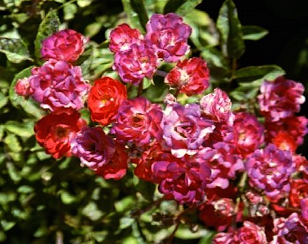
We may need to rethink some of the orange Polyanthas that “blue” to give an odd effect. For instance, ‘Golden Salmon’:
And to a lesser degree, ‘Orange Cameo’:
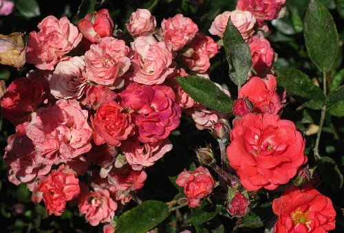
However, the picture of ‘Golden Salmon’ was taken Aug 7, 2000, ‘Orange Cameo’ on May 26, 2008, both in San Jose. The Polyanthas seem to have more of the purplish shading in Summer, and are clearer orange in cool Autumn weather.
But to recap, pelargonidin does a grey shade when bound to tannin spherules. In carnations, it gives a blue-grey color in AVIs (anthocyanic vacuolar inclusions). Stoddard’s cross of Orangeade x Angel Face suggests that a pelargonidin-based rosacyanin is also grey.
Now we need to learn how peonidin changes with tannin-spherules, AVIs and whether it can be the basis for a rosacyanin.
There is still more to be learned. Among other things, there can be an interaction between pigment concentration and solubility with cell shape. Yasuda discussed this in three papers:
http://bulbnrose.x10.mx/KKing/RosePigments/YasudaInsoluble.html
It may be that Rosa foetida is not the only species that contributed to the lilac color of ‘Grey Pearl’, ‘Sterling Silver’ and so one.
I’ve been reading about the colors and color patterns of sweet peas. A strain called ‘Blue Shift’ caught my attention because the blooms start out a purply pink then change to about the truest blue yet seen in the species. I had read that some of the various co-pigments are enhanced when connected with sugars, just like the anthocyanin pigments. I wondered whether a co-pigment responsible for turning the color to blue might be affected by a version of the “chameleon gene” like the one works in ‘Mutabilis’ and other changeable roses.
So, I went searching for more info on co-pigments in roses and promptly came up a paper: Profile of the Phenolic Compounds of Rosa rugosa Petals.
“Twenty polyphenols were identified by UPLC-ESI-MS. The main fraction of polyphenols was ellagitannins, which are 69 to 74% of the total polyphenols of the petals.”
This looks promising, because the rosacyanins have an “ellagitannin moiety”, according to Fukui et al (2006).
And as it happens, I have another obscure note reported by J. H. Nicolas (A Rose Odyssey, 1937):
In 1933 I had found a curious sport on Margaret McGredy. The foliage strongly resembled Rugosa but the plant characteristics also leaned toward R. cinnamomea. I mentioned this fact to Sam III [McGredy] when I visited him in 1934. Sam could not account for the sport. He had never used species in his breeding. His brother-in-law, Walter I. Johnston, spoke up, “Your father did much more work with species.” We adjourned to the office, where complete hybridizing records from the early days of the firm are kept, one volume for each year, a valuable library. After several hours of research we traced the origin of Margaret McGredy to crosses of Rugosa and Cinnamomea. They were, of course, many generations back. But as these two species are in the blood stream of Margaret McGredy and all modern McGredy roses, the possibility of the sport was explained."
This does not prove my case, but it may be that Rugosa has something to do with the appearance of Rosacyanins in a rose raised by Sam III.
It is possible that Rosa foetida did contribute in a useful way with a trait I’ve been calling “dominant non-red”. At the Heritage Rose Garden in San Jose, CA, I saw an alleged "Harison’s Yellow’ developing a red flush following a heatwave. This occurred in June 2003 and again in June 2006.
http://bulbnrose.x10.mx/Roses/Rose_Pictures/H/harisonsyellow.html
And just today I learned that ‘Agnes’ can produce apricot-pink blooms following a heatwave.
I don’t know the biochemistry of this trait, but it may be similar to the “chameleon trait” responsible for the pink or red color that appears after the flower is open. One difference, however, is that it blocks pigment formation before the bloom opens.
If this hypothetical gene is responsible for attaching a glucose molecule to the (otherwise colorless) cyanidin skeleton, but only functions at high temperatures, then at lower temps it would leave all the colorless cyanidin to be processed into rosacyanins before the bloom opens. If the “chameleon gene” is also present, whatever cyanidin is left would be the basis for the reddish border that develops on ‘Angel Face’, ‘Paradise’ and such.
Has anyone seen a mauve rose opening red following a heatwave?
BTW, ‘Sue Lawley’ is a “hand-painted” rose that seems to lose the effect at lower temperatures. However, I do have a picture of it looking quite ordinary in July.
http://bulbnrose.x10.mx/Roses/Rose_Pictures/S/suelawley.html
PS. Looks like my brain skipped a track. In order for the “dominant non-red” trait to be dominant, it is not enough to assume that it is a heat-sensitive version of a glucosyltransferase (to attach a glucose molecule or two to the cyanidin skeleton). It must block the action of the glucosyltransferase enzyme also present. This gets a bit stickier because roses have two types of glucosyltransferase: the rose glucosyltransferase RhGT1 catalyses two reactions instead of one, producing the common 3,5-glucosides. This is another example of how special roses really are. Then there is the 3-glucosyltransferase found in many plants besides roses.
A special form of this 3-glucosyltransferase is responsible for the chameleon trait. This is active only after the flower is open, so it would not conflict with the action of my hypothetical “dominant non-red” trait. Rugosas are colored mostly by 3,5-diglucosides, so it may be that the hypothetical gene somehow blocks RhGT1 [to account for ‘Agnes’] but not “special” version of 3-GT that acts after anthesis [for ‘Angel Face’, et al.]
Can rosacyanins co-exist with cyanidin-3-monoglucoside produced before opening? This would involve competition for the colorless cyanidin precursor. Eugster and Marki-Fisher (1991) mention three varieties that have more 3-monogluside than 3,5-diglucoside: ‘Souvenir de la Malmaison’, ‘François Juranville’ and ‘Dorothy Perkins’. In case anyone wants to aim for a pinker shade of mauve.
It occurred to me this morning that the ruby border on some mauve roses suggests that the production of rosacyanins can be limited by the availability of gallic acid. When the gallic acid is used up, any remaining leucocyanidin could be converted to chrysanthemin, if the light-sensitive form of 3-GT (glucosyltransferase) is present.
And on a possibly related note, many years ago I saw ‘Hidcote Gold’ with a few scattered mauve spots on some of the lower leaves … just where one might expect to see blackspot. At the time I guessed that the plant was pushing pigment and related substances into the infected areas to fight the fungus. But that was a long time before I learned about rosacyanins.
I did not get pictures, sorry to say. But I do recall that the lilac spots were uniform (not blotchy) and just the color of ‘Paradise’ or ‘Angel Face’.
I first saw ‘Smokey’ in Topeka. The color was interesting, but I never saw the bush produce a well-formed flower that opened properly. When I grew my own in Mission Viejo, it looked like a more terra cotta version of ‘Tropicana’.
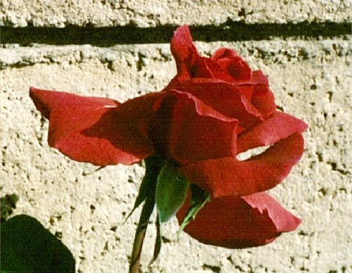
BTW, ‘Smokey’ was what I was aiming for when I crossed Sweet Chariot x Margo Koster. I liked what I got even though it was nowhere near ‘Smokey’.
